Investigational Treatments for Epidermolysis Bullosa:
A New Paradigm Focusing on Secondary Pathology
1 Introduction
Epidermolysis Bullosa (EB) is a heterogeneous group of rare, inherited, and currently incurable genetically determined by extreme mechanical fragility of the skin and mucous membranes, leading to blistering. Gloablly, there are approximately 500,000 people in the world living with EB (EB Research Partnership), with an incidence of 1.4–25.0 per million live births and a prevalence of 2.82–54.0 per million population (Baardman, et al.). The latest classification of EB identifies 4 major types based on the plane of ultrastructural fragility and blister formation, which reflects the different protein abnormality: EB simplex (EBS), junctional EB (JEB), dystrophic EB (DEB), and kindler EB (KEB) (Has, et al.). In each type of EB, the severe degree has been determined by the extent of cutaneous and extracutaneous involvement, protein expression levels, and specific genetic mutations.
1.1 EB Simplex
EB simplex is the most common type of EB accounting for about 70% of all EB (Bardhan, et al.) which defined by skin blistering due to cleavage within the basal layer of keratinocytes (Has, et al.). Clinically, EBS represents with intraepidermal blistering, ranging from localized palm involvement to severe generalized forms with clustered blisters everywhere on the surface of human body. It is primarily caused by the mutations in genes encoding keratin 5 (KRT5) and keratin 14 (KRT14), which disrupt the cytoskeletal in basal keratinocytes. These mutations will disrupt the structural proteins grouped as type I or type II keratins that form obligate heterodimers, which assemble through several stages into intermediate filaments and 3D cytoskeletons (Coulombe, et al.).
1.2 Junctional EB
Junctional EB results from autosomal recessive mutations in genes encoding lamina lucida within the basement membrane zone, such as laminin 332 (LAMA3, LAMB3, LAMC2) and type XVII collagen (COL17A1) (Bardhan, et al.). These defects impair the adhesion of basal keratinocytes to the underlying dermis, forming blister within the lamina lucida. In phenotype, it displays as generalized blistering, exuberant granulation tissue (particularly periorificial), enamel hypoplasia, and most severely high infant mortality.
1.3 Dystrophic EB
Dystrophic EB is characterized by cleavage in the upper dermis, arising in all cases from COL7A1 mutations that result in mutant type VII collagen and disrupted anchoring fibrils (Bardhan, et al.). Specifically, the mutations of gene COL7A1 will lead to the lack of VII collagen production, and finally end up with blistering in lamina densa, the deep level in between basal keratinocytes and the connective tissues. Two inheritance patterns, autosomal dominant or recessive, have different phenotypes. The recessive severe form is characterized by widespread blistering at birth, progressive mutilating scarring, and a profoundly elevated lifetime risk of aggressive squamous cell carcinoma arising in chronic wounds which can cause squamous carcinoma cancer.
1.4 Kindler EB
Kindler EB is a rare autosomal recessive type of EB which caused by the mutations in the FERMT1 gene (Jobard, et al.), encoding kindlin-1, a focal adhesion protein involved in integrin activtio nand keratinocyte adhesion. The blistering caused by the mutation of FERMT1 gene can occur at different levels of the dermal–epidermal junction — within the basal keratinocyte, along the lamina lucida or below the lamina densa of the epidermal basement membrane (Bardhan, et al.).
Recent research show that people have a relatively comprehensive understanding on the mechanisms of EB and have been continuously finding the fundamental solutions at the genetic level or the supportive management for patients. However, emerging therapeutic strategies are shifting to target the secondary pathological processes—such as chronic inflammation, fibrosis, impaired wound healing, and tumorigenesis—that drive disease progression and morbidity. This review will explore the available treatments for the secondary pathology of EB, with a specific explanation on the downstream pathological mechanisms and find the possible medicines to control or modulate these pathologies rather than solely correct the underlying genetic errors.
2 Current Challenges in Gene and Protein-Based Therapies
Although the inheritance pattern of EB includes autosomal dominant and autosomal recessive, dominant mutations often missense lead to dominant-negative interference. As a result, most of the investigation about gene therapies of EB focused on replacing genes in recessive forms of EB and silencing genes in dominant forms (Hou, et al.). The protein therapy has only been tried with DEB in a even smaller scale. Beyond this fundamental gene difficulty, several technical challenges have hindered the clinical translation of gene and protein-based approach.
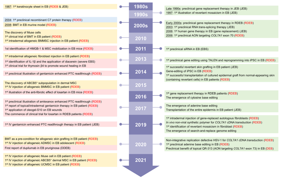
Fig. 1: A brief history of time for the evolution of EB-related therapies. This timeline highlights the milestone and the advancement of multiple treatment modalities for EB. (Hou, et al.)
2.1 Gene Approach
Gene approaches that have been tried in correcting EB-related gene mutations are gene replacement, gene editing, RNA-based, and revertant mosaicism. Gene replacement is a process of transduce the full-length wildtype cDNA to restore the gene expression and the function of a protein. Remarkably, Hirsch et al. used a 4 cm2 skin biopsy to derive correct LAMB3 sheets to a 7-year-old boy with intermediate JEB and treated 80% total body surface area (0.85 m2) (Hirsch, et al.). Plus, primary RDEB keratinocytes were transduced with full-length COL7A1 cDNA and cultured epidermal autografts were transplanted back to the wounds of four RDEB patients, 90% of the skin grafts showed new type VII collagen, although efficacy generally declined after 1 year (Eichstadt, et al.). By using gene replacement, the short-term of the lack proteins can be resolved efficiently, but in long-term, making these genes stay not silently is a big issue.
Editing genomes including inserting, deleting, modifying and replacing DNA, is a basic technique to probably heal the EB from the root with the support of recent-developed technologies. The emergence of gene editing for EB began in 2013, when transcription activator like effector nucleases (TALEN) were used to edit primary RDEB fibroblasts through homology-directed repair (Osborn, et al.). Since then, CRISPR-Cas9 has been introduced. This technique has applied in correcting pathogenetic mutations in EB-associated genes, including COL7A1, KRT14, and PLEC1(Hirsch, et al.; Murauer, et al.; Wally, et al. "K14 Mrna Reprogramming for Dominant Epidermolysis Bullosa Simplex"; Wally, et al. "5' Trans-Splicing Repair of the Plec1 Gene"). This approach aims for restoring the functional errors present in the protein expression and improving skin. Though gene editing is a transformative strategy to achieve long-term or root correction for EB, no gene editing methods have entered the clinical trials and all of them remain great uncertainties at the implementation level.
2.2 Protein Approach
In most of the recessive EB, the mutation on the genes will lead to the reduction or the complete absence of protein production. Therefore, one therapeutic option is to immediately give the recombinant protein to the abnormal cells, restoring skin anchoring structure. This protein approach has been used in treating C7 in RDEB and laminin-332 in JEB (Hou, et al.). For instance, intravenously administering recombinant C7 into the skin with RDEB has been shown to localize dermal-epidermal junction, promote the formation of anchoring fiber, finally enhancing wound healing in animal model (Woodley, et al.). Similarly, protein-based strategies may also include engineered variants with improved stability or delivery profiles. While protein replacement can offer a direct and rapid therapy for mitigating the structural deficits in EB, challenges remain in immunogenicity and tissue targeting.
3 Downstream Pathologies Driving Disease Burden
Beyond the disorders on EB patients’ skin, the mutation of genes triggers to severe symptoms, a series of irrelated downstream pathological process that are responsible for profound disease burden. Targeting these secondary pathways is the most viable near-term strategy to alleviate suffering through all the subtypes of EB and potentially alter the disease course.
3.1 Pain and Pruritic
Pain and itch are universal features for almost all EB patients, their pathophysiology go beyond the wounds, involving intricate neuro-immune crosstalk. For pain, the severe level varies among different type of EB: Severe pain is reported in 14% of patients with all types of epidermolysis bullosa, RDEB is the epidermolysis bullosa type that most commonly presents with pain, with at least 50% of patients suffering intense pain daily, and only 5% of patients reporting to be pain-free (Fine, et al.). Studies have confirmed that even patients with RDEB which do not have open wounds also exhibit small-fiber neuropathy determined by the reduce of intraepidermal nerve fiber density and abnormal pain perception (von Bischhoffshausen, et al.). This discovery established a clear neuropathological substrate for chronic pain of EB: the continuous tissue damage lead to the release of chemical signals like NGF and cytokines, making the hypersensitize pain-sensing nerves. Then, sensitization happened, the sensitized nerves send amplified pain signals to the spinal cord that’s experiencing maladaptive changes. This is why patients always feel severe pain from even a light touch and the pain persists beyond the wounds. (von Bischhoffshausen, et al.)
For pruritic, research confirmed that keratin in EBS directly stress the keratinocyte, causing the release of Thymic Stromal Lymphopoietin (TSLP), which is a powerful pruritogen. 8 of 17 patients with EBS and most of the keratin-deficient mice show elevated TSLP serum level, providing a high correlation between the TSLP level and the pruritic level (Kumar, et al.). Then the rising level of TSLP activate the cytokine receptors like IL-4, IL-13, and IL-31, which are known to be involved in itch pathway. After releasing, TSLP can bind to receptors on the nerve endings, amplifying pruritic signaling. (Papanikolaou, et al.)
3.2 Inflammation
Inflammation on EB patients is a cell-autonomous process initiated by the gene mutations and the tissue damage. It is confirmed that the inflammatory process of EBS involve multiple signaling pathways. Key pathway is the c-Jun N-terminal kinase (JNK) stress that activated by pro-inflammatory cytokines like IL-1b and IL-6. These cytokines are significantly increased in EBS patients (Lu, et al.) and promote the transcription of gene encoding matrix-degrading proteins like KLK5 and KLK7, fostering skin destabilization indeed. In addition, IFN-γ signaling play a role in inflammation by promoting signal transducer and activator of transcription (STAT) 1 phosphorylation and K17 stressing response. The one of the major activated pathways in EBS, phosphatidylinositol 3-kinase (PI3K)-protein kinase B (Akt)-mTOR pathway, is activated in EBS-blistered epidermis with upstream regulators including TNF-a, IL-1b, IL-2, IL-6, PI3K, and mTOR. Furthermore, dysregulation of wnt-receptor signaling pathway and bone morphogenetic proteins signaling (BMPs) may also link inflammation to increase the abnormal skin development in EBS.(Bchetnia, et al.) Finally, the T helper type 17 (Th17) immune response, marked by elevated level of IL-17, IL-21, and IL-22 in blister tissues, is significantly increased in severe EBS (Castela, et al.). This indicate a sustained inflammatory state that create obstacles for skin repairment. Conclusively, these all signaling pathway are potential imflammatory mechanisms that agrevate the skin fragility in EBS patients.
3.3 Fibrosis and Cancer
Fibrosis and squamous cell carcinoma (SCC) are two primary causes of death in RDEB and some JEB. These two complications share the same central molecular driver, the transforming growth factor-beta (TGF-b). Normal type of VII collagen will provide physical anchorage and regulatory signaling. Once VII collagen absence, the Thrombospondin-1 (TSP1) will accumulate in the extracellular matrix (ECM), which is a major activator of latent TGF-b (Atanasova, et al.). Once TGF-b activated, it forms a pro-fibrotic environment, ultimately leading to fibrotic diseases (Massague, and Sheppard). Critically, abnormal TGF-b signaling fundamentally reprograms the dermal microenvironment from tumor-suppressive to tumor-promoting (Pickup, et al.). This change in environment initiate, invade and metastasized by inducing expression of αvβ6 integrin in keratinocytes, which then activate more TGF-b (Zambruno, et al.), and promoting epithelial-mesenchymal transition (EMT) and induce angiogenesis (G. Han, et al.).
4 Targeting Secondary Pathologies with Reproposed and Novel Therapeutics
Despite the advantages of using gene therapies, resolve the root cause of EB and completely remove the symptoms for patients, implementing them is an extremely complicated and hard process. Think outside the box, heal the secondary pathologies can greatly improve the quality of life of patients. Several medicines have been proposed to treat pain, pruritic, chronic inflammation, fibrosis and the SCC.
4.1 Targeting pain and pruritic
4.1.1 Tetrahydrocannabinol and Cannabidiol
The endocannabinoid system, a group of endogenous cannabinoid receptors located in the central (CB1) and peripheral nervous system (CB2), can regulate pain. In EB, 3 case studies show that combining tetrahydrocannabinol (THC) and cannabidiol (CBD) oral spray control the pain in RBED patients (Schrader, et al.). THC is a partial agonist of CB1/2 and has been shown to stimulate b-endorphin production (Pertwee), which kills the pain, and CBD is able to antagonize undesired effects of THC such as sedation and intoxication while concurrently improving desirable effects like analgesia (Russo). This combination may be useful in disrupting the scratch-pain cycle.
4.1.2 Gabapentin and Pregabalin
Gabapentin and pregabalin are structurally related compounds with recognized efficacy in the treatment of neuropathic pain (Sills). They bind to the α2δ subunit of voltage-gated calcium channels, selectively reducing the excitatory neurotransmitter reduce (Sills). This match with the neuropathic pain component validated in RDEB and may be effective in directly addressing the pain. Remarkably, a trial of pregabalin treatment for RDEB pain and itch has been completed and proved that this medicine significantly reduced mean pain scores in 10 patients (Calvo, et al.).
4.1.3 Dupilumab
Dupilumab is a fully humanized monoclonal antibody targeting the interleukin-4 receptor alpha (IL-4Ra), a receptor subunit shared by two type 2 cytokines, IL-4 and IL-13, which play an important role in the pathogenesis of atopic dermatitis (Shehadeh, et al.). By inhibiting IL-4 or IL-13 signaling, dupilumab can reduce the production of TSLP, the pruritogenic cytokines. Then, the reduced amount of pruritogenic cytokines will restrain the immune activation and subsequent neural sensitization and finally reduce the feel of itch for patients.
4.2 Dampening chronic inflammation
4.2.1 Apremilast
Phosphodiesterase-4 (PDE4), mainly present in immune cells, epithelial cells, and brain cells, manifests as an intracellular non-receptor enzyme that normally breaks down cyclic adenosine monophosphate (cAMP), modulating inflammation and epithelial integrity (Li, et al.). As mentioned above in severe EBS patients, a stronger Th17 inflammatory signature is shown in their skin. Apremilast, and anti-Th17 molecule, increases intracellular cAMP by inhibiting PDE4 (AMGEN). A case study has explained that the apremilast treatment reduce the blistering and inflammation in EBS patients caused by the Th17 signaling pathway (Castela, et al.).
4.3 Modulating fibrosis and pro-tumor environment
4.3.1 Losartan
Losartan, a small-molecule angiotensin II type 1 receptor antagonist used to treat hypertension, was shown to reduce TGF-b expression and myocardial fibrosis in mice with hypertrophic (Lim, et al.). This is a potential medicine for disease-modifying candidate for RDEB. In Lim’s discovery, loartan has two function: reduce the blood presure and interfers with TGF-b signaling and production (Lim, et al.), which is the basic factor that drives fibrosis and SCC progression. In RDEB mouse models, losartan largely reduce the fibrosis and delay the tumor growth (Nystrom, et al.). It is possible that losartan can alter the natural mechanism of RDEB by lightening the pro-fibrotic and pro-tumorigenic microenvirenment. A phase I/II clinical trial is processing to evaluate the safeting of this treatment in RDEB patients (European Medicines Agency).
4.3.2 5-Fluorouracil (5-FU)
5-FU works by irreversibly inhibiting thymidylate synthase, disrupting DNA and RNA synthesis and then suppressing the rapid proliferation of cancer cells. It is common to use it in the treatment of basal-cell carcinoma and actinic keratosis dermatologically at a low dose, about 0.5%, and in combination with salicylic acid, which can boost the drug's absorption through the epidermis and contributing to its keratolytic impact. (S. N. Han, et al.)
5 Discussion
This review discusses a different perspective in EB management, from viewing it as a genetic fragility disorder to targeting its downstream pathologies, pain and itch, chronic inflammation and fibrosis and cancer. This aspect allows the clinical treatment that transform existing drugs like losartan, apremilast and dupilumab to a viable tool to resolve secondary pathologies of EB. This is significant in reducing patients’ most pressing symptoms and potentially become the bridge to future gene therapies. Though this approach is direct and promising, it faces several key challenges. Questions of optimal timing, the combination of therapies and the side effect remain, making implementing into more clinical trials even harder. Ultimately, focusing on these downstream pathways cannot replace the genetic methods but creates an actionable way to reduce suffering for patients today. This marks an undeniable evolution process from passive support to proactive support, reserving more power and hope in the hands of patients during the continue search for the genetic therapies.
6 Bibliography
AMGEN. “Mechanism of Action (MOA) | Otezla® (Apremilast) HCP.” Otezla® (Apremilast) Healthcare Professional Site, 2023, www.otezlapro.com/how-otezla-works/.
Atanasova, V. S. et al. "Thrombospondin-1 Is a Major Activator of Tgf-Beta Signaling in Recessive Dystrophic Epidermolysis Bullosa Fibroblasts." J Invest Dermatol, vol. 139, no. 7, 2019, pp. 1497-505 e5, Medline, doi:10.1016/j.jid.2019.01.011.
Baardman, R. et al. "Novel Insights into the Epidemiology of Epidermolysis Bullosa (Eb) from the Dutch Eb Registry: Eb More Common Than Previously Assumed?" J Eur Acad Dermatol Venereol, vol. 35, no. 4, 2021, pp. 995-1006, Medline, doi:10.1111/jdv.17012.
Bardhan, A. et al. "Epidermolysis Bullosa." Nat Rev Dis Primers, vol. 6, no. 1, 2020, p. 78, Medline, doi:10.1038/s41572-020-0210-0.
Bchetnia, M. et al. "Pathological Mechanisms Involved in Epidermolysis Bullosa Simplex: Current Knowledge and Therapeutic Perspectives." Int J Mol Sci, vol. 25, no. 17, 2024, Medline, doi:10.3390/ijms25179495.
Calvo, M. et al. "Pregabalin for Neuropathic Pain and Itch in Recessive Dystrophic Epidermolysis Bullosa: A Randomized Crossover Trial." JAMA Dermatol, vol. 160, no. 12, 2024, pp. 1314-19, Medline, doi:10.1001/jamadermatol.2024.3767.
Castela, E. et al. "Epidermolysis Bullosa Simplex Generalized Severe Induces a T Helper 17 Response and Is Improved by Apremilast Treatment." Br J Dermatol, vol. 180, no. 2, 2019, pp. 357-64, Medline, doi:10.1111/bjd.16897.
Coulombe, P. A. et al. "Epidermolysis Bullosa Simplex: A Paradigm for Disorders of Tissue Fragility." J Clin Invest, vol. 119, no. 7, 2009, pp. 1784-93, Medline, doi:10.1172/JCI38177.
EB Research Partnership. “What Is EB?” EB Research Partnership, 2023, ebresearch.org/what-is-eb.html. Accessed 29 Dec. 2025.
Eichstadt, S. et al. "Phase 1/2a Clinical Trial of Gene-Corrected Autologous Cell Therapy for Recessive Dystrophic Epidermolysis Bullosa." JCI Insight, vol. 4, no. 19, 2019, Medline, doi:10.1172/jci.insight.130554.
European Medicines Agency. “EudraCT Number 2015-003670-32 - Clinical Trial Results - EU Clinical Trials Register.” Clinicaltrialsregister.eu, 2015, www.clinicaltrialsregister.eu/ctr-search/trial/2015-003670-32/results. Accessed 31 Dec. 2025.
Fine, J. D. et al. "Assessment of Mobility, Activities and Pain in Different Subtypes of Epidermolysis Bullosa." Clin Exp Dermatol, vol. 29, no. 2, 2004, pp. 122-7, Medline, doi:10.1111/j.1365-2230.2004.01428.x.
Han, G. et al. "Distinct Mechanisms of Tgf-Beta1-Mediated Epithelial-to-Mesenchymal Transition and Metastasis during Skin Carcinogenesis." J Clin Invest, vol. 115, no. 7, 2005, pp. 1714-23, Medline, doi:10.1172/JCI24399.
Han, S. N. et al. "Clinical Treatment Study of Secondary Multiple Squamous Cell Carcinoma with Psoriasis Vulgaris." J Oncol, vol. 2022, 2022, p. 9529681, PubMed-not-MEDLINE, doi:10.1155/2022/9529681.
Has, C. et al. "Consensus Reclassification of Inherited Epidermolysis Bullosa and Other Disorders with Skin Fragility." Br J Dermatol, vol. 183, no. 4, 2020, pp. 614-27, Medline, doi:10.1111/bjd.18921.
Hirsch, T. et al. "Regeneration of the Entire Human Epidermis Using Transgenic Stem Cells." Nature, vol. 551, no. 7680, 2017, pp. 327-32, Medline, doi:10.1038/nature24487.
Hou, P. C. et al. "Investigational Treatments for Epidermolysis Bullosa." Am J Clin Dermatol, vol. 22, no. 6, 2021, pp. 801-17, Medline, doi:10.1007/s40257-021-00626-3.
Jobard, F. et al. "Identification of Mutations in a New Gene Encoding a Ferm Family Protein with a Pleckstrin Homology Domain in Kindler Syndrome." Hum Mol Genet, vol. 12, no. 8, 2003, pp. 925-35, Medline, doi:10.1093/hmg/ddg097.
Kumar, V. et al. "Keratin-Dependent Thymic Stromal Lymphopoietin Expression Suggests a Link between Skin Blistering and Atopic Disease." J Allergy Clin Immunol, vol. 138, no. 5, 2016, pp. 1461-64 e6, Medline, doi:10.1016/j.jaci.2016.04.046.
Li, H. et al. "Phosphodiesterase-4 Inhibitors for the Treatment of Inflammatory Diseases." Front Pharmacol, vol. 9, 2018, p. 1048, PubMed-not-MEDLINE, doi:10.3389/fphar.2018.01048.
Lim, D. S. et al. "Angiotensin Ii Blockade Reverses Myocardial Fibrosis in a Transgenic Mouse Model of Human Hypertrophic Cardiomyopathy." Circulation, vol. 103, no. 6, 2001, pp. 789-91, Medline, doi:10.1161/01.cir.103.6.789.
Lu, H. et al. "Induction of Inflammatory Cytokines by a Keratin Mutation and Their Repression by a Small Molecule in a Mouse Model for Ebs." J Invest Dermatol, vol. 127, no. 12, 2007, pp. 2781-9, Medline, doi:10.1038/sj.jid.5700918.
Massague, J. and D. Sheppard. "Tgf-Beta Signaling in Health and Disease." Cell, vol. 186, no. 19, 2023, pp. 4007-37, Medline, doi:10.1016/j.cell.2023.07.036.
Murauer, E. M. et al. "Functional Correction of Type Vii Collagen Expression in Dystrophic Epidermolysis Bullosa." J Invest Dermatol, vol. 131, no. 1, 2011, pp. 74-83, Medline, doi:10.1038/jid.2010.249.
Nystrom, A. et al. "Losartan Ameliorates Dystrophic Epidermolysis Bullosa and Uncovers New Disease Mechanisms." EMBO Mol Med, vol. 7, no. 9, 2015, pp. 1211-28, Medline, doi:10.15252/emmm.201505061.
Osborn, M. J. et al. "Talen-Based Gene Correction for Epidermolysis Bullosa." Mol Ther, vol. 21, no. 6, 2013, pp. 1151-9, Medline, doi:10.1038/mt.2013.56.
Papanikolaou, M. et al. "Prevalence, Pathophysiology and Management of Itch in Epidermolysis Bullosa." Br J Dermatol, vol. 184, no. 5, 2021, pp. 816-25, Medline, doi:10.1111/bjd.19496.
Pertwee, R. G. "The Diverse Cb1 and Cb2 Receptor Pharmacology of Three Plant Cannabinoids: Delta9-Tetrahydrocannabinol, Cannabidiol and Delta9-Tetrahydrocannabivarin." Br J Pharmacol, vol. 153, no. 2, 2008, pp. 199-215, Medline, doi:10.1038/sj.bjp.0707442.
Pickup, M. et al. "The Roles of Tgfbeta in the Tumour Microenvironment." Nat Rev Cancer, vol. 13, no. 11, 2013, pp. 788-99, Medline, doi:10.1038/nrc3603.
Russo, E. B. "Cannabinoids in the Management of Difficult to Treat Pain." Ther Clin Risk Manag, vol. 4, no. 1, 2008, pp. 245-59, PubMed-not-MEDLINE, doi:10.2147/tcrm.s1928.
Schrader, N. H. B. et al. "Combined Tetrahydrocannabinol and Cannabidiol to Treat Pain in Epidermolysis Bullosa: A Report of Three Cases." Br J Dermatol, vol. 180, no. 4, 2019, pp. 922-24, Medline, doi:10.1111/bjd.17341.
Shehadeh, W. et al. "Treatment of Epidermolysis Bullosa Pruriginosa-Associated Pruritus with Dupilumab." Br J Dermatol, vol. 182, no. 6, 2020, pp. 1495-97, Medline, doi:10.1111/bjd.18855.
Sills, G. J. "The Mechanisms of Action of Gabapentin and Pregabalin." Curr Opin Pharmacol, vol. 6, no. 1, 2006, pp. 108-13, Medline, doi:10.1016/j.coph.2005.11.003.
von Bischhoffshausen, S. et al. "Recessive Dystrophic Epidermolysis Bullosa Results in Painful Small Fibre Neuropathy." Brain, vol. 140, no. 5, 2017, pp. 1238-51, Medline, doi:10.1093/brain/awx069.
Wally, V. et al. "K14 Mrna Reprogramming for Dominant Epidermolysis Bullosa Simplex." Hum Mol Genet, vol. 19, no. 23, 2010, pp. 4715-25, Medline, doi:10.1093/hmg/ddq405.
Wally, V. et al. "5' Trans-Splicing Repair of the Plec1 Gene." J Invest Dermatol, vol. 128, no. 3, 2008, pp. 568-74, Medline, doi:10.1038/sj.jid.5701152.
Woodley, D. T. et al. "Intravenously Injected Recombinant Human Type Vii Collagen Homes to Skin Wounds and Restores Skin Integrity of Dystrophic Epidermolysis Bullosa." J Invest Dermatol, vol. 133, no. 7, 2013, pp. 1910-3, Medline, doi:10.1038/jid.2013.10.
Zambruno, G. et al. "Transforming Growth Factor-Beta 1 Modulates Beta 1 and Beta 5 Integrin Receptors and Induces the De Novo Expression of the Alpha V Beta 6 Heterodimer in Normal Human Keratinocytes: Implications for Wound Healing." J Cell Biol, vol. 129, no. 3, 1995, pp. 853-65, Medline, doi:10.1083/jcb.129.3.853.